To Track Your Order
Learn About Eye Care
3) Dry Eyes & Dry Eye Syndrome
7) Am I Entitled to FREE Eye Care?
Macular Degeneration
What is Macular Degeneration?
Age-related macular degeneration (ARMD) is the commonest cause of vision loss in people aged over 50 years old. The prevalence (number of new cases each year) increases with age. It is caused by degeneration of the macula, the central and most sensitive part of the retina at the back of the eye.
What is the Macula?
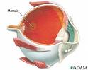 The macula is the central part of the retina which is responsible for enabling fine detail to be discerned. The remainder of the retina enables 'peripheral vision' only. Without the use of the macula, tasks like reading small print and recognising faces become difficult or impossible. The macula contains a yellow pigment (hence the term macula lutea).
The disease becomes increasingly more common amongst people in their 60s and 70s. By the age 75, almost 15% of people have this condition to some extent. The biggest risk factor is thus age. Other risk factors are a family history of the condition, cigarette smoking, and being white caucasian.
What are the types of ARMD?
There are two main types of ARMD often termed 'Dry ARMD' and 'Wet ARMD'. The pathological process is different between the two. In the wet form, there is a proliferation of abnormal blood vessels under the macula. In dry ARMD, there is the collection of small yellow deposits within the retina called drusen, and degeneration (atrophy) of the retinal tissue at the macula. The dry form is more common, but the wet form is usually more sudden and devastating to the vision.
| Photograph showing typical dry ARMD | Photograph of the macula showing multiple drusen | Photograph of the macula showing 'Wet' ARMD |
The paler elevated area at the macula represents the area where the retina is elevated, under which there is an abnormal 'membrane' due to abnormal proliferation of blood vessels.
What Does ARMD Do To The Vision?
ARMD affects only the central area of the vision. The condition thus never causes complete blindness or loss of sight.
| Normal Vision (Left) | Blurred Vision in ARMD (right) |
Is there any treatment for ARMD?
Currently, there is no cure for ARMD. The risk of developing ARMD can be reduced by not smoking. Studies have given us some evidence that a diet rich in antioxidants and certain pigments (found in dark green vegetables like broccoli and kale) may reduce the risk of progression of the disease process.
For a very small percentage of 'wet' ARMD cases, a treatment called 'Photo Dynamic Therapy' (PDT) may be used to reduce the risk of further visual deterioration.
LUCENTIS Injections
There is now a NICE approved treatment for wet ARMD called LUCENTIS. This is known as an 'Anti-VEGF' agent. VEGF is an acronym and stands for Vascular Endothelial Growth Factor. Lucentis is able to reduce the proliferation of the abnormal blood vessels that grow under the retina in wet ARMD. The drug is administered by injetion into the eye. A minimum of 3 injections are required, with the average number of injections required being 7.
LUCENTIS has been shown to significantly improve the final visual outcomes for a large proprtion of patients with wet ARMD. However, the treatment only works in the early stages of disease onset, and is not effective once the wet ARMD has caused chronic scarring of the retina.
Back to Top